Fiscal 2004
(1) Discovery of the "Raman Spectroscopic Signature of Life" in a Living Fission Yeast Cell
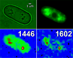
In a previous study, we found an intense unknown Raman band at 1602 cm-1 in the mitochondria of a living fission yeast cell (Schizosaccharomyces pombe). From a Raman mapping experiment of a cell whose mitochondria are GFP labeled (Fig. 1), we proved that the 1602 cm-1 band came exclusively from mitochondria. We also found that the intensity of this band was correlated strongly with the living activity of the cell. This relationship of the 1602 cm-1 band with the cell activity was examined by a respiration inhibiting experiment using KCN. Soon after adding KCN, the 1602 cm-1 band became weaker, while the other Raman bands ascribed to phospholipids remained unchanged. Then, the 1602 cm-1 band became further weaker and disappeared eventually, and the phospholipid bands changed their shapes gradually. We suspect that the intensity of the 1602 cm-1 band is a measure of the metabolic activity in mitochondria and that the gradual changes of the phospholipid bands arise from the structural degradation of the mitochondrial membranes induced by the lowered metabolic activity. These Raman spectral changes are most likely to reflect the early dying process of a living yeast cell. In this regard, we call the 1602 cm-1 band the "Raman spectroscopic signature of life". The discovery of this signature has opened up a new possibility of molecular level elucidation and quantification of life.

Fig. 1. Microscopic image (upper left), GFP image of mitochondria (upper right) and Raman mapping images of a fission yeast cell. The 1446 cm-1 band is from phospholipids.
(2) Development of Infrared Electroabsorption (IREA) Spectroscopy and Its Application to Structural Studies
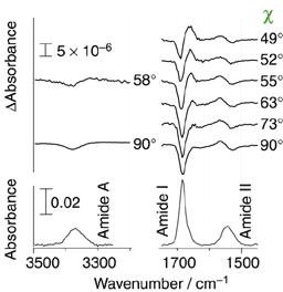
Infrared electroabsorption (IREA) spectroscopy provides unique higher-order vibrational information on molecular species existing in liquids and solutions. Species having distinct dipole moments are detected distinctly by IREA spectra and their dipole moments are determined separately. On account of technical difficulties, however, IREA was so far applicable only to wavenumber regions higher than 3000 cm-1. We developed a new IREA system based on a dispersive spectrometer and the AC-coupling detection scheme. It is capable of detecting infrared absorbance changes as small as 10-8 in ΔA and enables the measurements of IREA in the fingerprint region. Fig. 2 shows the IREA spectra of a 1,4-dioxane solution of N-methylacetamide observed with varying angle χ (the angle between the incident infrared electric vector and the direction of the applied external electric field. The high S/N ratio in these spectra enabled us to analyze the χ dependence by SVD (Singular Value Decomposition) and determine the dipole moments of the dimer as well as the monomer of N-methylacetamide in 1,4-dioxane. The analysis shows that the dimmer has a head-to-tail structure. As far as we are aware, this is the first experimental structural determination of a dimmer species in solution.

Fig. 2. Infrared electroabsorption spectrum of N-methylacetamide in 1,4-dioxane.
Refereed Journals
- Carrier Dynamics in TiO2 and Pt/TiO2 Powders Observed by Femtosecond Time-Resolved Near-Infrared Spectroscopy at a Spectral Region of 0.9-1.5 ƒÊm with the Direct Absorption Method. Koichi Iwata, Tomohisa Takaya, Hiro-o Hamaguchi, Akira Yamakata, Taka-aki Ishibashi, Hiroshi Onishi, and Haruo Kuroda,J. Physical Chem. B, 108, 52 20233-20239 (2004).
- Femtosecond coherent anti-Stokes Raman scattering spectroscopy using supercontinuum generated from a photonic crystal fiber. Hideaki Kano and Hiro-o Hamaguchi, Appl. Phys. Lett., 85, 19 4298-4300 (2004).
- Femtosecond electron transfer dynamics of 9,9'-bianthryl in acetonitrile as studied by time-resolved near-infraredabsorption spectroscopy. Tomohisa Takaya, Hiro-o Hamagushi, Haruo Kuroda, Koichi Iwata, Chem.Phys.Lett., 399, 210-214 (2004).
- Intercalation-induced structural change of DNA as studied by 1064 nm near-infrared multichannel Raman spectroscopy. Kenro Yuzaki and Hiro-o Hamaguchi, J. Raman Spectrosc., 35, 1013-1015 (2004).
- Discovery of a Magnetic Ionic Liquid [bmim]FeCl4. Satoshi Hayashi and Hiro-o Hamaguchi, Chem. Lett. 33, 12 1590-1591 (2004).
- Structure of an ionic liquid, 1-n-butyl-3-methylimidazolium iodide, studied by wide-angle X-ray scattering and Raman spectroscopy. Hideaki Katayanagi, Satoshi Hayashi, Hiro-o Hamaguchi and Keiko Nishikawa, Chem. Phys. Lett., 392, 460-464 (2004).
- Raman spectroscopic signature of life in a living yeast cell. Yu-San Huang, Takeshi Karashima, MasayukiYamamoto, Takashi Ogura and Hiro-o Hamaguchi, J. Raman Spectrosc., 35, 525-526 (2004).
- Orientational ordering of alkyl chain at the air/liquid interface of ionic liquids studied by sum frequency vibrational spectroscopy. Toshifumi Iimori, Takashi Iwahashi, Hisao Ishii, Kazuhiko Seki, Yukio Ouchi,Ryosuke Ozawa, Hiro-o Hamaguchi and Doseok Kim, Chem. Phys. Lett., 389, 321-326 (2004).
- Development of Infrared Electroabsorption Spectroscopy and Its Application to Molecular Structural Studies. Hirotsugu Hiramatsu and Hiro-o Hamaguchi, Appl. Spectrosc., 58, 4 355-366 (2004).