
Direct Raman Imaging Microspectroscopic System
Raman imaging of living cells
- visualizes molecular distributions in living cells.
- enables pursuit of molecular dynamics in vivo.
- So far...
- Scanning confocal Raman microscope
→ long measurement time, a few to a few tens of minutes.
→ unable to image the whole cell simultaneously - Present study...
- Direct Raman imaging microscope
→ shorter measurement time, a few tens of seconds
→ enable to image the whole cell simultaneously
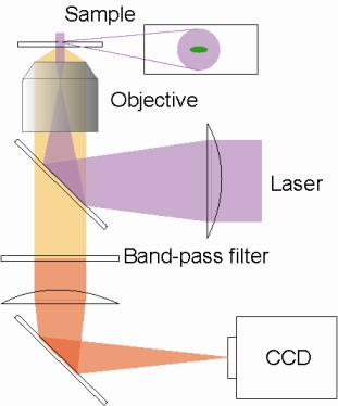
Fig. 1. Principle of direct Raman imaging method
Laser beam is expanded at the sample point. Raman scattered light is separated by a band-pass filter and imaged on a CCD detector directly.
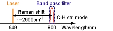
Fig. 2. Separation of Raman scattered light by a band-pass filter
Experimental Setup
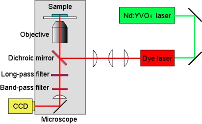
Fig. 3. Setup
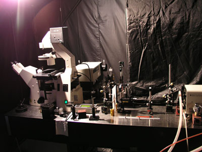
Fig. 4. Experimental Setup
Images of a fission yeast cell
Exposure time: 30 sec
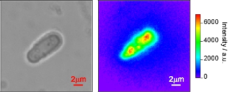
FIg. 5. Optical image, and 649nm excitation (∼2900cm-1). Raman image with C-H stretch modes visualizes clearly not only the shape of the cell but also mitochondria which are rich in C-H bonds.